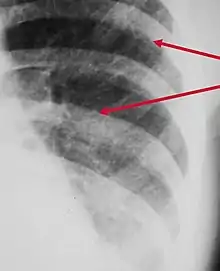
Peribronchial cuffing
Peribronchial cuffing, also referred to as peribronchial thickening or bronchial wall thickening, is a radiologic sign which occurs when excess fluid or mucus buildup in the small airway passages of the lung causes localized patches of atelectasis (lung collapse).[1] This causes the area around the bronchus to appear more prominent on an X-ray. It has also been described as donut sign, considering the edge is thicker, and the center contains air.
Examples
Peribronchial cuffing is seen in a number of conditions including:
- Acute bronchitis
- Asthma following exercise or during an acute episode
- Bronchiolitis
- Bronchopulmonary dysplasia
- Congestive heart failure
- Cystic fibrosis
- Diffuse parenchymal lung disease
- Extreme exertion through physical exercise
- Hantavirus pulmonary syndrome
- Human metapneumovirus
- Kawasaki disease
- Lung cancer
- Pneumonia
- Pulmonary edema
- Smoke inhalation
Treatment
As peribronchial cuffing is a sign rather than a symptom or condition, there is no specific treatment except to treat the underlying cause.
References
- Bramson RT, Griscom NT, Cleveland RH (2005). "Interpretation of chest radiographs in infants with cough and fever". Radiology. 236 (1): 22–29. doi:10.1148/radiol.2361041278. PMID 15983074.
This article is issued from Wikipedia. The text is licensed under Creative Commons - Attribution - Sharealike. Additional terms may apply for the media files.