X
wikiHow is a “wiki,” similar to Wikipedia, which means that many of our articles are co-written by multiple authors. To create this article, 26 people, some anonymous, worked to edit and improve it over time.
There are 7 references cited in this article, which can be found at the bottom of the page.
This article has been viewed 166,333 times.
Learn more...
Prokaryotes and eukaryotes are terms used to define types of organisms. The main difference between the two is the presence of a “true” nucleus: eukaryotes have one, while prokaryotes do not. Although this is the most easily recognizable difference, there are other important distinctions between the two organisms that can be seen under a microscope.
Steps
Part 1
Part 1 of 2:
Using a Microscope
-
1Obtain a specimen slide. Specimen slides of prokaryotes and eukaryotes can be obtained from biological supply companies.
- If you are in school, ask your science teacher if they have access to slides.
-
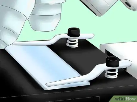
2Place your specimen slide on the microscope stage (the platform that holds the slides).[1] Some microscopes will have stage clips that allow you to secure the slide in place to prevent movement during focusing and viewing. If there are clips on the stage, gently push the slide underneath to secure in place. If no clips are provided, place the slide directly under objective.
- Be careful when pushing slides underneath the clips. Too much force can damage the slide.
- You may have to move the slide while looking through the eyepiece to find the desired area of the specimen.
Advertisement -
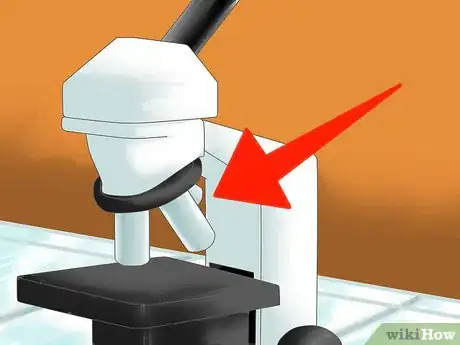
3Ensure the microscope is on the lowest magnification. The part of the microscope that allows for magnification is called an objective. Objectives for compound light microscopes usually range from 4x to 40x. You can move to higher magnifications if necessary, but starting low allows you to easily find the specimen on the slide.[2]
- You can determine the magnification of the objective by looking at the objective itself where it will have a label.
- The lowest magnification objective will also have the shortest length, while the highest magnification will have the longest length.
-
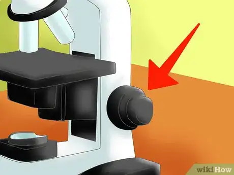
4Focus the image. Looking at a blurry image makes it difficult to make out small structures and define aspects of the cell. To more clearly see every detail, make sure the image is in focus.[3]
- While looking into the eyepiece, use the focus adjustment knobs found below the stage on the side of the microscope.
- By turning the knobs, you will see the image come into sharper focus.
-
5Increase the magnification if necessary. At the lowest magnification, you might notice that it's difficult to see smaller features and cellular structures. By increasing the magnification, you will be able to see more details within the cell.[4]
- Never change the objective while looking through the eyepiece. Because higher magnification objectives have longer lengths, changing the objective before lowering the stage can result in damage to the slide, the objective, and the microscope itself.
- Use the focus knobs to lower the stage to the appropriate height.
- Shift the objectives until the desired magnification is over the slide.
- Refocus the image.
Advertisement
Part 2
Part 2 of 2:
Observing the Image
-
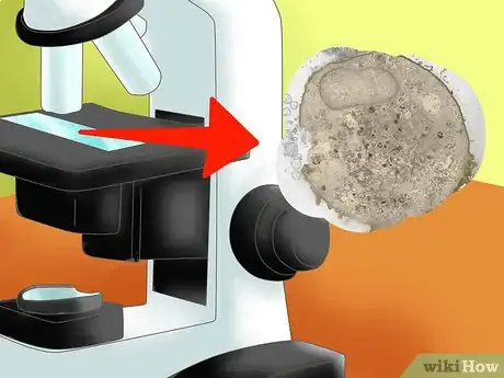
1Identify the features of eukaryotes. Eukaryotic cells are large and have many structural and internal components. The word eukaryote is rooted in the Greek language. "Káruon" means "kernel" which refers to the nucleus, while "eû" means "true" so eukaryotes contain a true nucleus. Eukaryotic cells are complex and contain membrane-bound organelles that perform specific functions to keep the cell alive.[5]
- Look for the nucleus of the cell.[6] The nucleus is the structure of a cell that contains the genetic information encoded by DNA. Although the DNA is linear, the nucleus generally appears as a dense circular mass inside cell.
- See if you can find organelles within the cytoplasm (the jelly-like interior of the cell). Under the microscope, you should be able to see distinct masses that are rounded or oblong in shape and smaller than the nucleus.
- All eukaryotes have a plasma membrane and cytoplasm, and some (plants and fungi) have a cell wall.[7] The plasma membrane will not be obvious under the microscope, but the cell wall should appear as a dark line outlining the edge of the cell.
- Although there are single-celled eukaryotes (protozoa), most are multi-cellular (animals and plants).
-
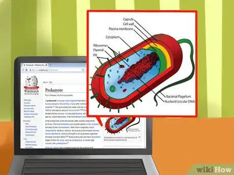
2Identify the features of prokaryotes. Prokaryotic cells are much smaller and have fewer internal structures. In Greek, "pró" means before, therefore prokaryote means "before a nucleus". Due to the absence of organelles they are simpler cells and perform fewer functions to stay alive.[8]
- Look for the absence of a nucleus. The genetic material of prokaryotes is not contained within a membrane-bound nucleus, instead, it floats around freely in the cytoplasm. The region where the genetic material resides is called the nucleoid, although this usually isn't visible under a regular microscope.[9]
- Other structures, such as ribosomes, are too small to see with a regular light microscope.[10]
- All prokaryotes have a cell membrane and cytoplasm, and most also have a cell wall. As with eukaryotic cells, the plasma membrane may not be obvious under the microscope, but the cell wall should be visible.
- Most prokaryotic cells are 10-100 times smaller than eukaryotic cells, although there are exceptions to this.
- All bacteria are prokaryotes. Example bacteria include Escherichia coli (E. coli), which lives in your intestines, and Staphylococcus aureus, which can cause skin infections.
-
3Observe the image through the microscope. Look through the microscope and write down the defining characteristics you see from the slide. Based on the specific features of eukaryotes and prokaryotes, you should be able to determine what is on your slide.
- Make a checklist for eukaryotes and prokaryotes and check off the features that apply to the specimen you're looking at.
Advertisement
Community Q&A
-
QuestionHow do I tell if a cell is prokaryotic or eukaryotic by looking at it?
 Community AnswerLook for the nucleus of the cell. Eukaryotes have a nucleus; prokaryotes don't.
 -
QuestionHow do I know if a cell is a eukaryotic?
 Community AnswerIf the sample given is a eukaryotic cell, then the compartments will be observed under a light microscope. In the case of proprokaryotes, no compartments are observed.
 -
QuestionDoes a virus have a prokaryotic or eukaryotic cell?
 Community AnswerNeither. Viruses are not cellular in nature, but instead are comprised of particles called virions. Instead of being contained by a cellular structure, viruses are enclosed by capsids.
Advertisement
References
- ↑ http://www.microscope-microscope.org/basic/microscope-parts.htm
- ↑ https://www2.mrc-lmb.cam.ac.uk/microscopes4schools/microscopes2.php
- ↑ https://www2.mrc-lmb.cam.ac.uk/microscopes4schools/microscopes2.php
- ↑ https://courses.lumenlearning.com/suny-mcc-microbiologylab2/chapter/use-of-the-microscope/
- ↑ https://opentextbc.ca/biology/chapter/3-2-comparing-prokaryotic-and-eukaryotic-cells/
- ↑ https://www.khanacademy.org/science/ap-biology/gene-expression-and-regulation/dna-and-rna-structure/a/prokaryote-structure
- ↑ https://www.khanacademy.org/science/ap-biology/gene-expression-and-regulation/dna-and-rna-structure/a/prokaryote-structure
- ↑ https://opentextbc.ca/biology/chapter/3-2-comparing-prokaryotic-and-eukaryotic-cells/
- ↑ https://www.livescience.com/65922-prokaryotic-vs-eukaryotic-cells.html
About This Article
Advertisement

