Podospora anserina
Podospora anserina is a filamentous ascomycete fungus from the order Sordariales. It is considered a model organism for the study of molecular biology of senescence (aging), prions, sexual reproduction, and meiotic drive.[1][2] It has an obligate sexual and pseudohomothallic life cycle. It is a non-pathogenic coprophilous fungus that colonizes the dung of herbivorous animals such as horses, rabbits, cows and sheep.[1][3]
| Podospora anserina | |
|---|---|
 | |
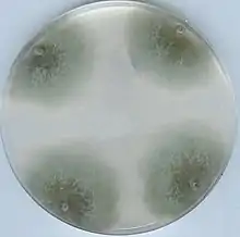
| Wild-type strain on a Petri dish | |
| Scientific classification | |
| Domain: | Eukaryota |
| Kingdom: | Fungi |
| Division: | Ascomycota |
| Class: | Sordariomycetes |
| Order: | Sordariales |
| Family: | Podosporaceae |
| Genus: | Podospora |
| Species: | P. anserina |
| Binomial name | |
| Podospora anserina | |
| Synonyms | |
Taxonomy
Podospora anserina was originally named Malinvernia anserina Rabenhorst (1857). Podospora anserina was subsequently published in Niessl (1883),[4] which is used today to reference the common laboratory strain therefrom, 'Niessl'. It is also known as Pleurage anserina (Ces.) Kuntze.[5][6] Genetics of P. anserina were characterized in Rizet and Engelmann (1949) and reviewed by Esser (1974). P. anserina is estimated to have diverged from Neurospora crassa 75 million years ago based on 18S rRNA and protein orthologous share 60-70% homology.[7] Gene cluster orthologs between Aspergillus nidulans and Podospora anserina have 63% identical primary amino acid sequence (even though these species are from distinct classes) and the average amino acid of compared proteomes is 10% less, giving rise to hypotheses of distinct species yet shared genes.
Research
Podospora is a model organism to study genetics, aging (senescence, cell degeneration), ascomycete development, heterokaryon incompatibility,[8] mating in fungi, prions, and mitochondrial and peroxisomal physiology.[9] Podospora is easily culturable on complex (full) potato dextrose, cornmeal agar/broth, or even synthetic medium, and, using modern molecular tools, is easy to manipulate. Its optimal growth temperature is 25–27 °C (77–81 °F) and can complete its life cycle in 7 to 11 days under laboratory conditions.[1][10]
Strains
Most research has been done in a small collection of French strains sampled in the 1920s, in particular the strains named S and s.[11] These two strains are known to be very similar except for the het-s locus. The reference genome published in 2008 corresponds to S+, a haploid derivate of the S strain with a + mating type.[7]
In addition, two other populations have been sampled, one in Usingen, Germany,[12] and the other in Wageningen, the Netherlands,[13][14][15][16] both of which have been used to study spore killing, the phenotypic expression of meiotic drive in fungi.[2]
In addition there are multiple lab-derived strains:
- ΔPaKu70 is used to increase homologous recombination in protoplasts during transformations in order to create desirable gene deletions or allelic mutations. A ΔPaKu70 strain can be achieved by transforming protoplasts with linear DNA that flanks the PaKu70 gene along with an antibiotic cassette and then selecting for strains and verifying by PCR. Derived from the s strain.[9]
- Mn19 is a long-lived strain used to study senescence. It is derived from strain A+-84-11 grown on manganese (Mn). This particular strain has been reported to have lived over 2 years in a race tube covering over 400 cm (160 in) of vegetative growth.[17]
- ΔiΔviv is an immortal strain that shows no sign of senescence. It produces yellow pigmentation. Lack of viv increased life span in days by a factor of 2.3 compared to the wild type and lack of i by 1.6. However, strain ΔiΔviv showed no senescence during the whole study and was vegetative for over a year. These genes are synergistic and are physically closely linked.[18]
- AL2 is a long-lived strain. Insertion of linear mitochondrial plasmid containing al-2 show increased life span. However, natural isolates that have homology to al-2 do not show increased life span.[19]
- Δgrisea is a long-lived strain and copper uptake mutant. This strain has lower affinity to copper and thus lower intracellular copper levels, leading to use of the cyanide-resistant alternative oxidase, PaAOX, pathway (instead of copper-dependent mitochondrial cytochrome c oxidase (COX) complex). This strain also exhibits more stable mtDNA. Copper use is similar to Δex1 strain.[20]
- Δex1 is an 'immortal strain' that has been grown for over 12 years and still shows no signs of senescence. This strain respires via a cyanide-resistant, salicylhydroxamic acid (SHAM)-sensitive pathway. This deletion disrupts the COX complex.[20]
Aging
Podospora anserina has a definite life span and shows senescence phenotypically (by slower growth, less aerial hyphae, and increased pigment production in distal hyphae). However, isolates show either increased life span or immortality, as to study the process of aging many genetic manipulations have been done to produce immortal strains or increase life-span. In general, the mitochondrion and mitochondrial chromosome is investigated.
Senescence occurs because during respiration reactive oxygen species are produced that limit the life span and over time defective mitochondrial DNA can accumulate.[19][21] With this knowledge, focus turned to nutrition availability, respiration (ATP synthesis) and oxidases, such as cytochrome c oxidase. Carotenoids, pigments that are also found in plants and provide health benefits to humans,[22] are known to be in fungi like Podospora's divergent ancestor Neurospora crassa; in N. crassa (and other fungi) cartenoids al genes namely provide UV radiation protection. Over-expression of al-2 Podospora anserina increased life span by 31%.[23]
Calorie restriction studies show that decreasing nutrients, such as sugar, increased life span, likely due to slower metabolism and thus decreased reactive oxygen species production or induced survival genes. Also, intracellular copper levels were found to be correlated with growth. This was studied in Grisea-deleted and ex1-deleted strains, as well as in a wild type s strain. Podospora without Grisea, a copper transcription factor, had decreased intracellular copper levels which lead to use of an alternative respiratory pathway that consequently produced less oxidative stress.[20]
Heterokaryon incompatibility
The following genes, both allelic and nonallelic, are found to be involved in vegetative incompatibility (only those cloned and characterized are listed): het-c, het-c, het-s, idi-2, idi-1, idi-3, mod-A, mode-D, mod-E, psp-A. Podospora anserina contains at least 9 het loci.[24]
Enzymes
Podospora anserina is known to produce laccases, a type of phenoloxidase.[25]
Genetics
Original genetic studies by gel electrophoresis led to the finding of the genome size, c. 35 megabases, with 7 chromosomes and 1 mitochondrial chromosome. In the 1980s the mitochondrial chromosome was sequenced. Then, in 2003, a pilot study was initiated to sequence regions bordering chromosome V's centromere using BAC clones and direct sequencing.[26] In 2008, a 10x whole genome draft sequence was published.[7] The genome size is now estimated to be 35-36 megabases.[7]
Genetic manipulation in fungi is difficult due to low homologous recombination efficiency and ectopic integrations[27] which hinders genetic studies using allele replacement and knock-outs.[9] In 2005, a method for gene deletion was developed based on a model for Aspergillus nidulans that involved cosmid plasmid transformation. A better system for Podospora was developed in 2008 by using a strain that lacked nonhomologous end joining proteins (Ku (protein), known in Podospora as PaKu70). This method claimed to have 100% of transformants undergo desired homologous recombination leading to allelic replacement, and after the transformation, the PaKu70 deletion can be restored by crossing over with a wild-type strain to yield progeny with only the targeted gene deletion or allelic exchange (e.g. point mutation).[9]
Secondary metabolites
It is well known that many organisms across all domains produce secondary metabolites, and fungi are prolific in this regard. Product mining was well underway in the 1990s for the genus Podospora. From Podospora anserina two new natural products classified as pentaketides, specifically derivatives of benzoquinones, were discovered; these showed antifungal, antibacterial, and cytotoxic activities.[28] Horizontal gene transfer is common in bacteria and between prokaryotes and eukaryotes yet is more rare between eukaryotic organisms. Between fungi, secondary metabolite clusters are good candidates for horizontal gene transfer, for example, a functional ST gene cluster that produces sterigmatocystin was found in Podospora anserina and originally derived from Aspergillus. This cluster is well-conserved, including, notably, the transcription-factor binding sites. Sterigmatocystin itself is toxic and is a precursor to another toxic metabolite, aflatoxin.[29]
See also
References
- Silar P (2020). Podospora anserina. France: HAL.
- Vogan AA, Martinossi-Allibert I, Ament-Velásquez SL, Svedberg J, Johannesson H (2022-01-02). "The spore killers, fungal meiotic driver elements". Mycologia. 114 (1): 1–23. doi:10.1080/00275514.2021.1994815. PMID 35138994. S2CID 246700229.
- Bills GF, Gloer JB, An Z (October 2013). "Coprophilous fungi: antibiotic discovery and functions in an underexplored arena of microbial defensive mutualism". Current Opinion in Microbiology. 16 (5): 549–565. doi:10.1016/j.mib.2013.08.001. PMID 23978412.
- Niessl von Mayendorf, Gustav (1883). "Ueber die Theilung der Gattung Sordaria" (PDF). Hedwigia (in German). 22: 153–156. Retrieved 9 July 2023.
- Torrey Botanical Club (2021) [1902]. Memoirs of the Torrey Botanical Club. Creative Media Partners, LLC. ISBN 978-1-01-357071-1.
- "Podospora anserina". Mycobank.
- Espagne E, Lespinet O, Malagnac F, Da Silva C, Jaillon O, Porcel BM, et al. (2008). "The genome sequence of the model ascomycete fungus Podospora anserina". Genome Biology. 9 (5): R77. doi:10.1186/gb-2008-9-5-r77. PMC 2441463. PMID 18460219.
- Bidard F, Clavé C, Saupe SJ (June 2013). "The transcriptional response to nonself in the fungus Podospora anserina". G3. 3 (6): 1015–1030. doi:10.1534/g3.113.006262. PMC 3689799. PMID 23589521.
- El-Khoury R, Sellem CH, Coppin E, Boivin A, Maas MF, Debuchy R, Sainsard-Chanet A (April 2008). "Gene deletion and allelic replacement in the filamentous fungus Podospora anserina". Current Genetics. 53 (4): 249–258. doi:10.1007/s00294-008-0180-3. PMID 18265986. S2CID 25538245.
- Vogan AA, Ament-Velásquez SL, Granger-Farbos A, Svedberg J, Bastiaans E, Debets AJ, et al. (July 2019). "Combinations of Spok genes create multiple meiotic drivers in Podospora". eLife. 8: e46454. doi:10.7554/eLife.46454. PMC 6660238. PMID 31347500.
- Rizet G (1952). "Les phénomènes de barrage chez Podospora anserina. I. Analyse genetique des barrages entre souches S et s." [The phenomena in Podospora anserina. I. Genetic analysis of barriers between S and s strains.]. Revue de cytologie et de biologie végétales; le botaniste. [Journal of Plant Cytology and Biology; the botanist] (in French). 13: 51–92.
- Hamann A, Osiewacz HD (December 2004). "Genetic analysis of spore killing in the filamentous ascomycete Podospora anserina". Fungal Genetics and Biology. 41 (12): 1088–1098. doi:10.1016/j.fgb.2004.08.008. PMID 15531213.
- Debets AJ, Dalstra HJ, Slakhorst M, Koopmanschap B, Hoekstra RF, Saupe SJ (June 2012). "High natural prevalence of a fungal prion". Proceedings of the National Academy of Sciences of the United States of America. 109 (26): 10432–10437. Bibcode:2012PNAS..10910432D. doi:10.1073/pnas.1205333109. PMC 3387057. PMID 22691498.
- Hermanns J, Debets F, Hoekstra R, Osiewacz HD (March 1995). "A novel family of linear plasmids with homology to plasmid pAL2-1 of Podospora anserina". Molecular & General Genetics. 246 (5): 638–647. doi:10.1007/BF00298971. PMID 7700237. S2CID 32936183.
- van der Gaag M, Debets AJ, Osiewacz HD, Hoekstra RF (June 1998). "The dynamics of pAL2-1 homologous linear plasmids in Podospora anserina". Molecular & General Genetics. 258 (5): 521–529. doi:10.1007/s004380050763. PMID 9669334. S2CID 23107862.
- van der Gaag M, Debets AJ, Oosterhof J, Slakhorst M, Thijssen JA, Hoekstra RF (October 2000). "Spore-killing meiotic drive factors in a natural population of the fungus Podospora anserina". Genetics. 156 (2): 593–605. doi:10.1093/genetics/156.2.593. PMC 1461285. PMID 11014809.
- Silliker ME, Cummings DJ (August 1990). "Genetic and molecular analysis of a long-lived strain of Podospora anserina". Genetics. 125 (4): 775–81. doi:10.1093/genetics/125.4.775. PMC 1204103. PMID 2397883.
- Esser K, Keller W (February 1976). "Genes inhibiting senescence in the ascomycete Podospora anserina". Molecular & General Genetics. 144 (1): 107–10. doi:10.1007/BF00277312. PMID 1264062. S2CID 8663226.
- Maas MF, de Boer HJ, Debets AJ, Hoekstra RF (September 2004). "The mitochondrial plasmid pAL2-1 reduces calorie restriction mediated life span extension in the filamentous fungus Podospora anserina". Fungal Genetics and Biology. 41 (9): 865–71. doi:10.1016/j.fgb.2004.04.007. PMID 15288022.
- Borghouts C, Werner A, Elthon T, Osiewacz HD (January 2001). "Copper-modulated gene expression and senescence in the filamentous fungus Podospora anserina". Molecular and Cellular Biology. 21 (2): 390–9. doi:10.1128/MCB.21.2.390-399.2001. PMC 86578. PMID 11134328.
- Pramanik D, Andasarie T, Özkan C (17 October 2012). "Genetic dissection of complex biological traits; the lifespan extending effect of calorie restriction in the filamentous fungus Podospora anserina". Code Groen.
- Johnson EJ (2002). "The role of carotenoids in human health". Nutrition in Clinical Care. 5 (2): 56–65. doi:10.1046/j.1523-5408.2002.00004.x. PMID 12134711.
- Strobel I, Breitenbach J, Scheckhuber CQ, Osiewacz HD, Sandmann G (April 2009). "Carotenoids and carotenogenic genes in Podospora anserina: engineering of the carotenoid composition extends the life span of the mycelium". Current Genetics. 55 (2): 175–84. doi:10.1007/s00294-009-0235-0. PMID 19277665. S2CID 32718690.
- Moore D, Frazer LN (June 2007). Essential Fungal Genetics. Springer Science & Business Media. p. 40. ISBN 978-0-387-22457-2.
- Esser K, Minuth W (1970). "The phenoloxidases of the ascomycete Podospora anserina. Communication 4. Genetic regulation of the formation of laccase". Genetics. 64 (3): 441–58. doi:10.1093/genetics/64.3-4.441. PMC 1212412. PMID 4988412.
- Silar P, Barreau C, Debuchy R, Kicka S, Turcq B, Sainsard-Chanet A, et al. (August 2003). "Characterization of the genomic organization of the region bordering the centromere of chromosome V of Podospora anserina by direct sequencing". Fungal Genetics and Biology. 39 (3): 250–263. doi:10.1016/s1087-1845(03)00025-2. PMID 12892638.
- Asch DK, Kinsey JA (March 1990). "Relationship of vector insert size to homologous integration during transformation of Neurospora crassa with the cloned am (GDH) gene". Molecular & General Genetics. 221 (1): 37–43. doi:10.1007/BF00280365. PMID 2157957. S2CID 24711141.
- Wang H, Gloer KB, Gloer JB, Scott JA, Malloch D (June 1997). "Anserinones A and B: new antifungal and antibacterial benzoquinones from the coprophilous fungus Podospora anserina". Journal of Natural Products. 60 (6): 629–31. doi:10.1021/np970071k. PMID 9214737.
- Slot JC, Rokas A (January 2011). "Horizontal transfer of a large and highly toxic secondary metabolic gene cluster between fungi". Current Biology. 21 (2): 134–139. doi:10.1016/j.cub.2010.12.020. PMID 21194949.
External links
 Media related to Podospora anserina at Wikimedia Commons
 Data related to Podospora anserina at Wikispecies