MALDI imaging
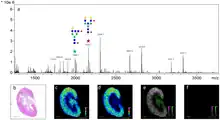
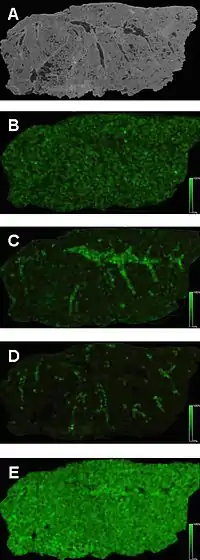
MALDI mass spectrometry imaging (MALDI-MSI) is the use of matrix-assisted laser desorption ionization as a mass spectrometry imaging[2] technique in which the sample, often a thin tissue section, is moved in two dimensions while the mass spectrum is recorded.[3] Advantages, like measuring the distribution of a large amount of analytes at one time without destroying the sample, make it a useful method in tissue-based study.[4]

Sample preparation

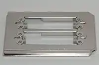
Sample preparation is a critical step in imaging spectroscopy. Scientists take thin tissue slices mounted on conductive microscope slides and apply a suitable MALDI matrix to the tissue, either manually or automatically. Next, the microscope slide is inserted into a MALDI mass spectrometer. The mass spectrometer records the spatial distribution of molecular species such as peptides, proteins or small molecules. Suitable image processing software can be used to import data from the mass spectrometer to allow visualization and comparison with the optical image of the sample. Recent work has also demonstrated the capacity to create three-dimensional molecular images using MALDI imaging technology and comparison of these image volumes to other imaging modalities such as magnetic resonance imaging (MRI).[5][6]
Tissue preparation
The tissue samples must be preserved quickly in order to reduce molecular degradation. The first step is to freeze the sample by wrapping the sample then submerging it in a cryogenic solution.[7] Once frozen, the samples can be stored below -80 °C for up to a year.[7] When ready to be analyzed, the tissue is embedded in a gelatin media which supports the tissue while it is being cut, while reducing contamination that is seen in optimal cutting temperature compound (OCT) techniques.[7][8][9] The mounted tissue section thickness varies depending on the tissue.
Tissue sections can then be thaw-mounted by placing the sample on the surface of a conductive slide that is of the same temperature, and then slowly warmed from below.[7] The section can also be adhered to the surface of a warm slide by slowly lowering the slide over the cold sample until the sample sticks to the surface.[7]
The sample can then be stained in order to easily target areas of interest, and pretreated with washing in order to remove species that suppress molecules of interest.[7][10] Washing with varying grades of ethanol removes lipids in tissues that have a high lipid concentration with little delocalization and maintains the integrity of the peptide spatial arrangement within the sample.[7][10][11]

Matrix application
The matrix must absorb at the laser wavelength and ionize the analyte. Matrix selection and solvent system relies heavily upon the analyte class desired in imaging. The analyte must be soluble in the solvent in order to mix and recrystallize the matrix. The matrix must have a homogeneous coating in order to increase sensitivity, intensity, and shot-to-shot reproducibility. Minimal solvent is used when applying the matrix in order to avoid delocalization.[13]
One technique is spraying. The matrix is sprayed, as very small droplets, onto the surface of the sample, allowed to dry, and re-coated until there is enough matrix to analyze the sample.[7] The size of the crystals depend on the solvent system used.
Sublimation can also be used to make uniform matrix coatings with very small crystals.[7][14] The matrix is placed in a sublimation chamber with the mounted tissue sample inverted above it.[7] Heat is applied to the matrix, causing it to sublime and condense onto the surface of the sample.[7] Controlling the heating time controls the thickness of the matrix on the sample and the size of the crystals formed.[7][14]
Automated spotters are also used by regularly spacing droplets throughout the tissue sample.[7] The image resolution relies on the spacing of the droplets.[7]
Applications
MALDI-MSI involves the visualization of the spatial distribution of proteins, peptides, lipids, and other small molecules within thin slices of tissue, such as animal or plant.[16][17][18][19] The application of this technique to biological studies has increased significantly since its introduction. MALDI-MSI is providing major contributions to the understanding of diseases, improving diagnostics, and drug delivery. Significant studies are of the eye, cancer research,[20] drug distribution,[21][22] and neuroscience.[9][23]
MALDI-MSI has been able to differentiate between drugs and metabolites[19] and provide histological information in cancer research, which makes it a promising tool for finding new protein biomarkers.[24][20][25] However, this can be challenging because of ion suppression, poor ionization, and low molecular weight matrix fragmentation effects. To combat this, chemical derivatization is used to improve detection.[26][27]
References
- Powers, Thomas W.; Neely, Benjamin A.; Shao, Yuan; Tang, Huiyuan; Troyer, Dean A.; Mehta, Anand S.; Haab, Brian B.; Drake, Richard R. (2014). "MALDI Imaging Mass Spectrometry Profiling of N-Glycans in Formalin-Fixed Paraffin Embedded Clinical Tissue Blocks and Tissue Microarrays". PLOS ONE. 9 (9): e106255. Bibcode:2014PLoSO...9j6255P. doi:10.1371/journal.pone.0106255. ISSN 1932-6203. PMC 4153616. PMID 25184632.
- McDonnell LA, Heeren RM (2007). "Imaging mass spectrometry" (PDF). Mass Spectrometry Reviews. 26 (4): 606–43. Bibcode:2007MSRv...26..606M. doi:10.1002/mas.20124. hdl:1874/26394. PMID 17471576.
- Chaurand P, Norris JL, Cornett DS, Mobley JA, Caprioli RM (2006). "New developments in profiling and imaging of proteins from tissue sections by MALDI mass spectrometry". J. Proteome Res. 5 (11): 2889–900. doi:10.1021/pr060346u. PMID 17081040.
- Walch, A.; Rauser, S.; Deininger, S. O.; Hofler, H. (2008). "MALDI imaging mass spectrometry for direct tissue analysis: a new frontier for molecular histology". Histochemistry and Cell Biology. 130 (3): 421–434. doi:10.1007/s00418-008-0469-9. PMC 2522327. PMID 18618129.
- Andersson M, Groseclose MR, Deutch AY, Caprioli RM (2008). "Imaging Mass Spectrometry of Proteins and Peptides: 3D Volume Reconstruction". Nature Methods. 5 (1): 101–108. doi:10.1038/nmeth1145. PMID 18165806. S2CID 29078650.
- Sinha TK, Khatib-Shahidi S, Yankeelov TE, Mapara K, Ehtesham M, Cornett DS, Dawant BM, Caprioli RM, Gore JC (2008). "Integrating Spatially Resolved Three-Dimensional MALDI IMS with in vivo Magnetic Resonance Imaging". Nature Methods. 5 (1): 57–59. doi:10.1038/nmeth1147. PMC 2649801. PMID 18084298.
- Norris, Jeremy L.; Caprioli, Richard M. (10 April 2013). "Analysis of Tissue Specimens by Matrix-Assisted Laser Desorption/Ionization Imaging Mass Spectrometry in Biological and Clinical Research". Chemical Reviews. 113 (4): 2309–2342. doi:10.1021/cr3004295. PMC 3624074. PMID 23394164.
- Cillero-Pastor, Berta; Heeren, Ron M. A. (7 February 2014). "Matrix-Assisted Laser Desorption Ionization Mass Spectrometry Imaging for Peptide and Protein Analyses: A Critical Review of On-Tissue Digestion". Journal of Proteome Research. 13 (2): 325–335. doi:10.1021/pr400743a. PMID 24087847.
- Chen, Ruibing; Hui, Limei; Sturm, Robert M.; Li, Lingjun (June 2009). "Three dimensional mapping of neuropeptides and lipids in crustacean brain by mass spectral imaging". Journal of the American Society for Mass Spectrometry. 20 (6): 1068–1077. doi:10.1016/j.jasms.2009.01.017. PMC 2756544. PMID 19264504.
- Chaurand, Pierre; Schwartz, Sarah A.; Billheimer, Dean; Xu, Baogang J.; Crecelius, Anna; Caprioli, Richard M. (February 2004). "Integrating Histology and Imaging Mass Spectrometry". Analytical Chemistry. 76 (4): 1145–1155. doi:10.1021/ac0351264. PMID 14961749.
- Hanrieder, J.; Ljungdahl, A.; Falth, M.; Mammo, S. E.; Bergquist, J.; Andersson, M. (6 July 2011). "L-DOPA-induced Dyskinesia is Associated with Regional Increase of Striatal Dynorphin Peptides as Elucidated by Imaging Mass Spectrometry". Molecular & Cellular Proteomics. 10 (10): M111.009308. doi:10.1074/mcp.M111.009308. PMC 3205869. PMID 21737418.
- Nilsson, Anna; Fehniger, Thomas E.; Gustavsson, Lena; Andersson, Malin; Kenne, Kerstin; Marko-Varga, György; Andrén, Per E.; Morty, Rory Edward (14 July 2010). "Fine Mapping the Spatial Distribution and Concentration of Unlabeled Drugs within Tissue Micro-Compartments Using Imaging Mass Spectrometry". PLOS ONE. 5 (7): e11411. Bibcode:2010PLoSO...511411N. doi:10.1371/journal.pone.0011411. PMC 2904372. PMID 20644728.
- Schwartz, Sarah A.; Reyzer, Michelle L.; Caprioli, Richard M. (July 2003). "Direct tissue analysis using matrix-assisted laser desorption/ionization mass spectrometry: practical aspects of sample preparation". Journal of Mass Spectrometry. 38 (7): 699–708. Bibcode:2003JMSp...38..699S. doi:10.1002/jms.505. PMID 12898649.
- Hankin, Joseph A.; Barkley, Robert M.; Murphy, Robert C. (September 2007). "Sublimation as a method of matrix application for mass spectrometric imaging". Journal of the American Society for Mass Spectrometry. 18 (9): 1646–1652. doi:10.1016/j.jasms.2007.06.010. PMC 2042488. PMID 17659880.
- Spraggins, Jeffrey M.; Caprioli, Richard M. (2011). "High-Speed MALDI-TOF Imaging Mass Spectrometry: Rapid Ion Image Acquisition and Considerations for Next Generation Instrumentation". J. Am. Soc. Mass Spectrom. 22 (6): 1022–1031. Bibcode:2011JASMS..22.1022S. doi:10.1007/s13361-011-0121-0. PMC 3514015. PMID 21953043.
- Caldwell RL, Caprioli RM (2005). "Tissue profiling by mass spectrometry: a review of methodology and applications". Mol. Cell. Proteomics. 4 (4): 394–401. doi:10.1074/mcp.R500006-MCP200. PMID 15677390.
- Reyzer ML, Caprioli RM (2007). "MALDI-MS-based imaging of small molecules and proteins in tissues". Current Opinion in Chemical Biology. 11 (1): 29–35. doi:10.1016/j.cbpa.2006.11.035. PMID 17185024.
- Woods AS, Jackson SN (2006). "Brain tissue lipidomics: direct probing using matrix-assisted laser desorption/ionization mass spectrometry". The AAPS Journal. 8 (2): E391–5. doi:10.1208/aapsj080244. PMC 3231574. PMID 16796390.
- Khatib-Shahidi S, Andersson M, Herman JL, Gillespie TA, Caprioli RM (2006). "Direct Molecular Analysis of Whole-body Animal Tissue Sections by Imaging MALDI Mass Spectrometry". Analytical Chemistry. 78 (18): 6448–6456. doi:10.1021/ac060788p. PMID 16970320.
- Rauser, Sandra; Marquardt, Claudio; Balluff, Benjamin; Deininger, Sören-Oliver; Albers, Christian; Belau, Eckhard; Hartmer, Ralf; Suckau, Detlev; Specht, Katja; Ebert, Matthias Philip; Schmitt, Manfred; Aubele, Michaela; Höfler, Heinz; Walch, Axel (5 April 2010). "Classification of HER2 Receptor Status in Breast Cancer Tissues by MALDI Imaging Mass Spectrometry". Journal of Proteome Research. 9 (4): 1854–1863. doi:10.1021/pr901008d. PMC 2918067. PMID 20170166.
- Khatib-Shahidi, Sheerin; Andersson, Malin; Herman, Jennifer L.; Gillespie, Todd A.; Caprioli, Richard M. (September 2006). "Direct Molecular Analysis of Whole-Body Animal Tissue Sections by Imaging MALDI Mass Spectrometry". Analytical Chemistry. 78 (18): 6448–6456. doi:10.1021/ac060788p. PMID 16970320.
- Acquadro, Elena; Cabella, Claudia; Ghiani, Simona; Miragoli, Luigi; Bucci, Enrico M.; Corpillo, Davide (April 2009). "Matrix-Assisted Laser Desorption Ionization Imaging Mass Spectrometry Detection of a Magnetic Resonance Imaging Contrast Agent in Mouse Liver". Analytical Chemistry. 81 (7): 2779–2784. doi:10.1021/ac900038y. PMID 19281170.
- Kutz, Kimberly K.; Schmidt, Joshua J.; Li, Lingjun (October 2004). "In Situ Tissue Analysis of Neuropeptides by MALDI FTMS In-Cell Accumulation". Analytical Chemistry. 76 (19): 5630–5640. doi:10.1021/ac049255b. PMID 15456280.
- Stoeckli M, Staab D, Schweitzer A (2006). "Compound and metabolite distribution measured by MALDI mass spectrometric imaging in whole-body tissue sections". International Journal of Mass Spectrometry. 260 (2–3): 195–202. Bibcode:2007IJMSp.260..195S. doi:10.1016/j.ijms.2006.10.007.
- Castellino, Stephen; Groseclose, M Reid; Wagner, David (November 2011). "MALDI imaging mass spectrometry: bridging biology and chemistry in drug development". Bioanalysis. 3 (21): 2427–2441. doi:10.4155/bio.11.232. PMID 22074284.
- Manier, M. Lisa; Reyzer, Michelle L.; Goh, Anne; Dartois, Veronique; Via, Laura E.; Barry, Clifton E.; Caprioli, Richard M. (13 May 2011). "Reagent Precoated Targets for Rapid In-Tissue Derivatization of the Anti-Tuberculosis Drug Isoniazid Followed by MALDI Imaging Mass Spectrometry". Journal of the American Society for Mass Spectrometry. 22 (8): 1409–1419. Bibcode:2011JASMS..22.1409M. doi:10.1007/s13361-011-0150-8. PMC 3424619. PMID 21953196.
- Franck, J.; El Ayed, M.; Wisztorski, M.; Salzet, M.; Fournier, I. (15 October 2009). "On-Tissue N-Terminal Peptide Derivatizations for Enhancing Protein Identification in MALDI Mass Spectrometric Imaging Strategies". Analytical Chemistry. 81 (20): 8305–8317. doi:10.1021/ac901043n. PMID 19775114.
Further reading
- Francese S, Dani FR, Traldi P, Mastrobuoni G, Pieraccini G, Moneti G (February 2009). "MALDI mass spectrometry imaging, from its origins up to today: the state of the art". Comb. Chem. High Throughput Screen. 12 (2): 156–74. doi:10.2174/138620709787315454. PMID 19199884.
- Richard M. Caprioli; Terry B. Farmer & Jocelyn Gile (December 1997). "Molecular Imaging of Biological Samples: Localization of Peptides and Proteins Using MALDI-TOF MS". Anal. Chem. 69 (23): 4751–4760. doi:10.1021/ac970888i. PMID 9406525.