C3orf67
Chromosome 3 open reading frame 67 or C3orf67 is a protein that in humans is encoded by the gene C3orf67.[5][6] The function of C3orf67 is not yet fully understood.
| CFAP20DC | |||||||||||||||||||||||||||||||||||||||||||||||||||
|---|---|---|---|---|---|---|---|---|---|---|---|---|---|---|---|---|---|---|---|---|---|---|---|---|---|---|---|---|---|---|---|---|---|---|---|---|---|---|---|---|---|---|---|---|---|---|---|---|---|---|---|
| Identifiers | |||||||||||||||||||||||||||||||||||||||||||||||||||
| Aliases | CFAP20DC, chromosome 3 open reading frame 67, C3orf67, CFAP20 domain containing | ||||||||||||||||||||||||||||||||||||||||||||||||||
| External IDs | MGI: 1926154 HomoloGene: 18873 GeneCards: CFAP20DC | ||||||||||||||||||||||||||||||||||||||||||||||||||
| |||||||||||||||||||||||||||||||||||||||||||||||||||
| |||||||||||||||||||||||||||||||||||||||||||||||||||
| |||||||||||||||||||||||||||||||||||||||||||||||||||
| |||||||||||||||||||||||||||||||||||||||||||||||||||
| Wikidata | |||||||||||||||||||||||||||||||||||||||||||||||||||
| |||||||||||||||||||||||||||||||||||||||||||||||||||
Gene
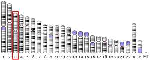
C3orf67 is located at 3p14.2 on the reverse strand ranging from 58716417 to 59050045 base pairs.[7][5] The accession number is NP_001338459.1.[8]
Protein
Primary sequence and isoforms
The coding sequence is 402-2681 base pairs of 3135 base pairs,[7] making up 759 amino acids.[5][8] C3orf67 has six validated isoforms.[5] Isoform one is the most complete with 16 exons.[7] C3orf67 weighs 84.35 kilodaltons.[9]
Post-translational modifications

Several post-translational modifications have been predicted for C3orf67 in conserved regions using various bioinformatic prediction tools[11][12][13][14][15][16][17][18]
- Two nuclear export signals
- Three sumoylation sites
- Two o-glycosylation sites
- One phosphorylation site
- One tyrosine sulfation site
Secondary structure
The beginning of C3orf67 is predicted to consist of a series of beta strands and a couple alpha helices that coincide with the DUF667 domain. There are also alpha helices predicted in regions that correspond to the CM_mono2 and OCRE domains.[19][20][21]
Tertiary structure

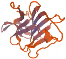
The DUF667 region is predicted to form a tube-like structure from a series of beta sheets.[21]
Homology and Evolution
Paralogs
There are no known paralogs of C3orf67.
Orthologs
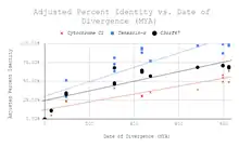
Orthologs have been identified for C3orf67 in species ranging from fungus, plants, hemichordates, parasites, fish, reptiles, birds, invertebrates, and mammals.
| Species | Common Name | Date of Divergence (MYA) | Accession Number | Sequence Length (aa) | % Identity |
| Orbicella faveolata | Mountainous star coral | 824 | XP_020630732.1 / XP_020630739.1 | 849 | 32.20% |
| Exaiptasia pallida | Pale anemone | 824 | XP_020899564.1 | 797 | 32.00% |
| Acanthaster planci | Crown-of-thorns starfish | 684 | XP_022107809.1 | 976 | 31.60% |
| Stylophora pistillata | Smooth cauliflower coral | 824 | XP_022782397.1 | 825 | 30.80% |
| Crassostrea gigas | Pacific oyster | 797 | XP_011453705.1 | 950 | 29.50% |
| Lingula anatina | Lamp shell | 797 | XP_013404893.1 | 1077 | 29.30% |
| Octopus bimaculoides | California two-spotted octopus | 797 | XP_014778712.1 | 902 | 29.10% |
| Saccoglossus kowalevskii | Acorn worm | 684 | XP_006821003.1 | 596 | 23.30% |
| Amphimedon queenslandica | Sponge | 951.8 | XP_011402616.1 | 508 | 22.70% |

Distant homologs
| Species | Common Name | Date of Divergence (MYA) | Accession Number | Sequence Length (aa) | % Identity |
| Trichinella spiralis | Trichina worm | 797 | XP_003374081.1 | 393 | 12.60% |
| Spizellomyces punctatus | Unknown | 1105 | XP_016608387.1 | 183 | 8.20% |
| Selaginella moellendorffii | Spikemoss | 1496 | XP_002989784.1 | 209 | 6.00% |
Expression
Promoter
The promoter is well conserved across humans, gibbons, baboons, orangutans, cats, squirrels, alpacas, rabbits and mice.[22] There are several high quality transcription factor binding sites.[23] There are also several stem-loop structures that are predicted to be formed in the promoter region, some of which overlap with transcription factor binding sites.[24]
Expression
C3orf67 is prominently expressed in the liver, tonsils, trachea, ovaries, testis, placenta, and colon. In other tissues it is expressed at low levels.[25] An increase in expression has been linked to small cell lung cancer.[26]
Function
The protein has been identified as one of seventeen (17) genes that may play a novel role in the intersection of tumor promotion and DNA-damaging stress and may be linked to carcinogenesis.[27]
Interacting Proteins
Transcription factors
There are three notable transcription factors that are known to be involved in the regulation of cell growth or immune responses:
Other interacting proteins
Several other proteins have been predicted to interact with C3orf67:
- CLK1[33]
- Phosphorylates serine/arginine-rich proteins involved in pre-mRNA processing in the nucleus.[34]
- CDK16 (gene)[35]
- A protein kinase thought to play a role in signal transduction cascades in differentiated cells, exocytosis, and transport of secretory cargo from the ER.[36]
- MARS2[37]
- Mitochondrial methionyl-tRNA synthetase.[38]
- AARS2[37]
- mitochondrial alanyl-tRNA synthetase.[38]
- C12orf60[39]
References
- GRCh38: Ensembl release 89: ENSG00000163689 - Ensembl, May 2017
- GRCm38: Ensembl release 89: ENSMUSG00000021747 - Ensembl, May 2017
- "Human PubMed Reference:". National Center for Biotechnology Information, U.S. National Library of Medicine.
- "Mouse PubMed Reference:". National Center for Biotechnology Information, U.S. National Library of Medicine.
- "NCBI Gene". National Center for Biotechnology Information.
- "C3orf67". GeneCards Human Gene Database.
- "NCBI Nucleotide". National Center for Biotechnology Information. May 2019.
- "NCBI Protein". National Center for Biotechnology Information.
- "Protein Molecular Weight Calculator". www.sciencegateway.org. Retrieved 2018-02-25.
- "MOTIF: Searching Protein Sequence Motifs". www.genome.jp. Retrieved 2018-02-25.
- "DictyOGlyc 1.1 Server". www.cbs.dtu.dk. Retrieved 2018-04-30.
- "GPS-SUMO: Prediction of SUMOylation Sites & SUMO-interaction Motifs". sumosp.biocuckoo.org. Retrieved 2018-04-30.
- "ExPASy - Sulfinator tool". web.expasy.org. Retrieved 2018-04-30.
- "SUMOplot™ Analysis Program | Abgent". www.abgent.com. Retrieved 2018-04-30.
- "C3orf67 (human)". www.phosphosite.org. Retrieved 2018-04-30.
- "NetOGlyc 4.0 Server". www.cbs.dtu.dk. Retrieved 2018-04-30.
- "YinOYang 1.2 Server". www.cbs.dtu.dk. Retrieved 2018-04-30.
- "NetPhos 3.1 Server". www.cbs.dtu.dk. Retrieved 2018-04-30.
- "JPred: A Protein Secondary Structure Prediction Server". www.compbio.dundee.ac.uk. Retrieved 2018-04-24.
- Kelley, Lawrence. "PHYRE2 Protein Fold Recognition Server". www.sbg.bio.ic.ac.uk. Retrieved 2018-04-24.
- "SWISS-MODEL | Workspace". swissmodel.expasy.org. Retrieved 2018-04-24.
- "Human BLAT Search". genome.ucsc.edu. Retrieved 2018-04-24.
- "Genomatix: Matrix Library information". www.genomatix.de. Retrieved 2018-04-24.
- "RNA Folding Form | mfold.rit.albany.edu". unafold.rna.albany.edu. Retrieved 2018-05-02.
- Dezso Z, Nikolsky Y, Sviridov E, Shi W, Serebriyskaya T, Dosymbekov D, Bugrim A, Rakhmatulin E, Brennan RJ, Guryanov A, Li K, Blake J, Samaha RR, Nikolskaya T (November 2008). "A comprehensive functional analysis of tissue specificity of human gene expression". BMC Biology. 6: 49. doi:10.1186/1741-7007-6-49. PMC 2645369. PMID 19014478.
- Sato T, Kaneda A, Tsuji S, Isagawa T, Yamamoto S, Fujita T, Yamanaka R, Tanaka Y, Nukiwa T, Marquez VE, Ishikawa Y, Ichinose M, Aburatani H (2013-05-29). "PRC2 overexpression and PRC2-target gene repression relating to poorer prognosis in small cell lung cancer". Scientific Reports. 3 (1): 1911. Bibcode:2013NatSR...3E1911S. doi:10.1038/srep01911. PMC 3665955. PMID 23714854.
- Glover KP, Chen Z, Markell LK, Han X (2 October 2015). "Synergistic Gene Expression Signature Observed in TK6 Cells upon Co-Exposure to UVC-Irradiation and Protein Kinase C-Activating Tumor Promoters". PLOS ONE. 10 (10): e0139850. Bibcode:2015PLoSO..1039850G. doi:10.1371/journal.pone.0139850. PMC 4592187. PMID 26431317.
- Zawel L, Dai JL, Buckhaults P, Zhou S, Kinzler KW, Vogelstein B, Kern SE (March 1998). "Human Smad3 and Smad4 are sequence-specific transcription activators". Molecular Cell. 1 (4): 611–7. doi:10.1016/S1097-2765(00)80061-1. PMID 9660945.
- "Genomatix: Matrix Library information". www.genomatix.de. Retrieved 2018-04-24.
- Treiber T, Mandel EM, Pott S, Györy I, Firner S, Liu ET, Grosschedl R (May 2010). "Early B cell factor 1 regulates B cell gene networks by activation, repression, and transcription- independent poising of chromatin". Immunity. 32 (5): 714–25. doi:10.1016/j.immuni.2010.04.013. PMID 20451411.
- "Genomatix: Matrix Library information". www.genomatix.de. Retrieved 2018-04-24.
- Molnár A, Georgopoulos K (December 1994). "The Ikaros gene encodes a family of functionally diverse zinc finger DNA-binding proteins". Molecular and Cellular Biology. 14 (12): 8292–303. doi:10.1128/MCB.14.12.8292. PMC 359368. PMID 7969165.
- Lipp JJ, Marvin MC, Shokat KM, Guthrie C (August 2015). "SR protein kinases promote splicing of nonconsensus introns". Nature Structural & Molecular Biology. 22 (8): 611–7. doi:10.1038/nsmb.3057. PMID 26167880. S2CID 24363149.
- "Antibodypedia - CLK1 antibodies". www.antibodypedia.com. Retrieved 2018-05-01.
- Mikolcevic P, Sigl R, Rauch V, Hess MW, Pfaller K, Barisic M, Pelliniemi LJ, Boesl M, Geley S (February 2012). "Cyclin-dependent kinase 16/PCTAIRE kinase 1 is activated by cyclin Y and is essential for spermatogenesis". Molecular and Cellular Biology. 32 (4): 868–79. doi:10.1128/MCB.06261-11. PMC 3272973. PMID 22184064.
- "Antibodypedia - CDK16 antibodies". www.antibodypedia.com. Retrieved 2018-05-01.
- van Meel E, Wegner DJ, Cliften P, Willing MC, White FV, Kornfeld S, Cole FS (October 2013). "Rare recessive loss-of-function methionyl-tRNA synthetase mutations presenting as a multi-organ phenotype". BMC Medical Genetics. 14: 106. doi:10.1186/1471-2350-14-106. PMC 3852179. PMID 24103465.
- "STRING: functional protein association networks". string-db.org. Retrieved 2018-05-01.
- Cornen S, Guille A, Adélaïde J, Addou-Klouche L, Finetti P, Saade MR, Manai M, Carbuccia N, Bekhouche I, Letessier A, Raynaud S, Charafe-Jauffret E, Jacquemier J, Spicuglia S, de The H, Viens P, Bertucci F, Birnbaum D, Chaffanet M (2014-01-09). "Candidate luminal B breast cancer genes identified by genome, gene expression and DNA methylation profiling". PLOS ONE. 9 (1): e81843. Bibcode:2014PLoSO...981843C. doi:10.1371/journal.pone.0081843. PMC 3886975. PMID 24416132.


