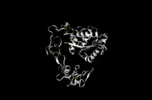
Atrolysin A
Atrolysin A (EC 3.4.24.1) is an enzyme that is one of six hemorrhagic toxins found in the venom of western diamondback rattlesnake. This endopeptidase has a length of 419 amino acid residues.[1][2][3][4] The metalloproteinase disintegrin-like domain and the cysteine-rich domain of the enzyme are responsible for the enzyme's hemorrhagic effects on organisms via inhibition of platelet aggregation.[5][6][7]
| Atrolysin A | |||||||||
|---|---|---|---|---|---|---|---|---|---|
| Identifiers | |||||||||
| EC no. | 3.4.24.1 | ||||||||
| CAS no. | 37288-82-7 | ||||||||
| Databases | |||||||||
| IntEnz | IntEnz view | ||||||||
| BRENDA | BRENDA entry | ||||||||
| ExPASy | NiceZyme view | ||||||||
| KEGG | KEGG entry | ||||||||
| MetaCyc | metabolic pathway | ||||||||
| PRIAM | profile | ||||||||
| PDB structures | RCSB PDB PDBe PDBsum | ||||||||
| |||||||||
| Zinc metalloproteinase-disintegrin-like atrolysin-A | |||||||
|---|---|---|---|---|---|---|---|
| Identifiers | |||||||
| Organism | |||||||
| Symbol | N/A | ||||||
| CAS number | 37288-82-7 | ||||||
| UniProt | Q92043 | ||||||
| Other data | |||||||
| EC number | 3.4.24.1 | ||||||
| |||||||
This enzyme catalyzes the following chemical reactions:
- cleavage of peptide bonds between amino acid residues Asn3-Gln4, His5-Leu6, His10-Leu11, Ala14-Leu15 and Tyr16-Leu17 in the insulin B chain
- removal of the C-terminal Leu from small peptides
Nomenclature
The accepted name for this enzyme is Atrolysin A. The enzyme is also known by:
- Crotalus atrox metalloendopeptidase a,
- Crotalus atrox α-proteinase,
- Crotalus atrox proteinase,
- bothropasin, and
- hemorrhagic toxin a
Structure
The amino acid sequence of atrolysin A is 419 sequences long, and contains ten calcium binding sites between positions 9 and 225, three of which are catalytic histidine residues at positions 142, 146, and 152.[6] There are also three positions (142, 146, 152) that are used for binding with the enzyme's cofactor, zinc. The active site lies at position 143.[6]

A significant portion of the protein from position 1-190 consists of alpha-helical structures with two shorted helixes between positions 315-340.[6] There are 39 observed disulfide bonds in the protein, all but one of these disulfide bonds occur between positions 157-409. Research suggests that the RSECD cysteine residue at positions 272-276 must be involved in a disulfide bond for the enzyme to have an inhibitory affect on platelet aggregation.[6]
The protein contains the following three domains:
- N-terminal Reprolysin (M12B) family zinc metalloprotease domain, followed by a
- disintegrin domain (containing a RSECD sequence analogous to RGD motif), and finally a
- C-terminal ADAM (A Disintegrin-like and Metalloproteinase) cysteine-rich domain.[8][5]
Mechanisms of action
The metalloproteinase disintegrin-like domain has shown an ability to degrade sub-endothelial matrix proteins such as type IV collagen, and fibronectin[6] which is partially responsible for the hemorrhage caused by the toxin
The cysteine-rich domain binds to the alpha-2/beta-1 integrin and causes inhibition of the platelet aggregation pathways that collagen is responsible for.[7][6] The glycoprotein VI (GPVI) collagen receptor however, does not seem affected by atrolysin A or other snake venom metalloproteinases (SVMP ).[9] Other agonists for platelet formation and aggregation such as adenosine diphosphate (ADP) or pathways mediated by convulxin also do not seem inhibited by the toxin in most organisms.[6][10][11] The cysteine-rich domain was shown to have two sequences that cause an interaction which prevents alpha-2/beta-1 integrin expressing K562 cells and platelets from binding to collagen.[9]
This enzyme may have the potential to inhibit platelet formation with certain pathways that were previously determined that the toxin had no effect on. For example, ADP stimulated aggregation appears to be inhibited by atrolysin A in a study where insect cells were used to express the protein, and the protein was inserted into human blood. When this variant of the protein was inserted into human blood, ADP stimulated platelet aggregation was inhibited.[5] This form of platelet aggregation does not appear inhibited by atrolysin A in other studies. This suggests that there may be an interaction between the disintegrin-like domain, and cysteine-rich domain of atrolysin A and fibrinogen receptor alpha-2b/beta-3 as well as the collagen receptor.[5]
Isozymes
Other atrolysin isozymes (A-F) have been further studied to understand potential methods of treatment against SVMPs. Research has been conducted on the use of medications to treat other diseases such as arthritis, and tumor metastasis as well.[12]
- Atrolysin B
- Atrolysin C
- Atrolysin D
- Atrolysin E
- Atrolysin F
References
- Bjarnason JB, Tu AT (August 1978). "Hemorrhagic toxins from Western diamondback rattlesnake (Crotalus atrox) venom: isolation and characterization of five toxins and the role of zinc in hemorrhagic toxin e". Biochemistry. 17 (16): 3395–3404. doi:10.1021/bi00609a033. PMID 210790.
- Mori N, Nikai T, Sugihara H, Tu AT (February 1987). "Biochemical characterization of hemorrhagic toxins with fibrinogenase activity isolated from Crotalus ruber ruber venom". Archives of Biochemistry and Biophysics. 253 (1): 108–121. doi:10.1016/0003-9861(87)90643-6. PMID 2949699.
- Bjarnason JB, Hamilton D, Fox JW (May 1988). "Studies on the mechanism of hemorrhage production by five proteolytic hemorrhagic toxins from Crotalus atrox venom". Biological Chemistry Hoppe-Seyler. 369 (Suppl): 121–129. PMID 3060135.
- Bjarnason JB, Fox JW (1989). "Hemorrhagic toxins from snake venoms". Journal of Toxicology: Toxin Reviews. 7 (2): 121–209. doi:10.3109/15569548809059729.
- Jia LG, Wang XM, Shannon JD, Bjarnason JB, Fox JW (May 1997). "Function of disintegrin-like/cysteine-rich domains of atrolysin A. Inhibition of platelet aggregation by recombinant protein and peptide antagonists". The Journal of Biological Chemistry. 272 (20): 13094–13102. doi:10.1074/jbc.272.20.13094. PMID 9148922.
- "Zinc metalloproteinase-disintegrin-like atrolysin-A - Crotalus atrox (Western diamondback rattlesnake)". www.uniprot.org. Retrieved 2021-10-29.
- Serrano SM, Jia LG, Wang D, Shannon JD, Fox JW (October 2005). "Function of the cysteine-rich domain of the haemorrhagic metalloproteinase atrolysin A: targeting adhesion proteins collagen I and von Willebrand factor". The Biochemical Journal. 391 (Pt 1): 69–76. doi:10.1042/BJ20050483. PMC 1237140. PMID 15929722.
- "Protein: VM3AA_CROAT (Q92043)". InterPro. European Molecular Biology Laboratory.
- Kamiguti AS, Gallagher P, Marcinkiewicz C, Theakston RD, Zuzel M, Fox JW (August 2003). "Identification of sites in the cysteine-rich domain of the class P-III snake venom metalloproteinases responsible for inhibition of platelet function". FEBS Letters. 549 (1–3): 129–134. doi:10.1016/S0014-5793(03)00799-3. PMID 12914938.
- Puri RN, Colman RW (1997). "ADP-induced platelet activation". Critical Reviews in Biochemistry and Molecular Biology. 32 (6): 437–502. doi:10.3109/10409239709082000. PMID 9444477.
- Francischetti IM, Ghazaleh FA, Reis RA, Carlini CR, Guimarães JA (May 1998). "Convulxin induces platelet activation by a tyrosine-kinase-dependent pathway and stimulates tyrosine phosphorylation of platelet proteins, including PLC gamma 2, independently of integrin alpha IIb beta 3". Archives of Biochemistry and Biophysics. 353 (2): 239–250. doi:10.1006/abbi.1998.0598. PMID 9606958.
- Zhang D, Botos I, Gomis-Rüth FX, Doll R, Blood C, Njoroge FG, et al. (August 1994). "Structural interaction of natural and synthetic inhibitors with the venom metalloproteinase, atrolysin C (form d)". Proceedings of the National Academy of Sciences of the United States of America. 91 (18): 8447–8451. doi:10.1073/pnas.91.18.8447. PMC 44623. PMID 8078901.
External links
- Atrolysin+A at the U.S. National Library of Medicine Medical Subject Headings (MeSH)